
Retina Glaucoma Center & Superspecialty Eye Hospital › Services › OCT Macula (optical coherence tomography)
Optical coherence tomography (OCT) provides cross-sectional images of the retina, the light-sensitive layer at the back of the eye that communicates with the brain and enables people to see. The macula is the part of the retina that allows central vision so people can see objects or text directly in front of them.
Our specialists are available 6 days a week for in-person and virtual consultations.
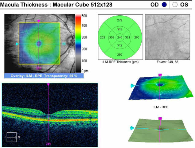
The macula is a tiny but extraordinarily important area at the centre of your retina. It is responsible for all your fine, detailed, and colour vision — everything you use when reading, recognising faces, watching television, or doing any close work. OCT Macula scanning produces incredibly detailed cross-sectional images of this vital area, allowing our specialists to detect changes and diagnose conditions that would be completely missed in a standard eye examination.
Optical Coherence Tomography uses light waves — in the same way that ultrasound uses sound waves — to produce high-resolution cross-sectional images of the retina. The macula scan focuses specifically on the central retina, capturing the thickness, structure, and integrity of each individual layer in extraordinary detail. The resulting images look almost like a microscopic slice through the retina, showing every layer clearly and allowing specialists to identify even very subtle abnormalities that would otherwise go undetected.
Dilating drops may be used to widen the pupils for a clearer and more complete view of the macula, though many modern OCT systems can perform high-quality scans without dilation.
You sit comfortably at the OCT machine and rest your chin on the support. You are asked to look at a small fixation target inside the machine and keep your eye as still as possible.
The machine scans the central retina automatically in a matter of seconds, capturing hundreds of cross-sectional images that are assembled into a detailed three-dimensional map of the macular tissue.
The images appear on the screen instantly. Your specialist reviews them immediately, identifies any abnormalities, compares them to normal values and previous scans, and discusses the findings with you clearly at the same appointment.
Completely safe and entirely comfortable. OCT uses light — not radiation of any kind — and is completely non-invasive. The scan is painless, takes only a few minutes, and has no aftereffects. It can be repeated as frequently as needed to monitor your condition over time without any risk to your eyes.
Need eye care? Contact us today.
East side of BRB Hospital, 57/E Panthapath, Dhaka-1215
Call Now