Retina Glaucoma Center & Superspecialty Eye Hospital › Services › FFA (Fundus Fluorescein Angiography)
Fundus fluorescein angiography (FFA) is an invasive diagnostic procedure. It helps to assess the anatomy, physiology, and pathology of retinal and choroidal circulation. It aids in the diagnosis of various ocular pathologies. It contributes to decision-making when planning the management of ocular pathology.
Our specialists are available 6 days a week for in-person and virtual consultations.
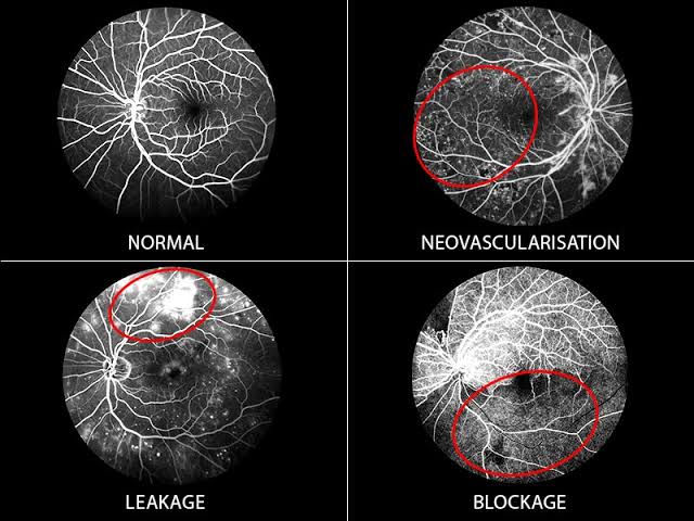
The tiny blood vessels of the retina can reveal a great deal about the health of your eye — but they are far too small and complex to examine properly with a standard eye check. FFA is a specialised imaging procedure that maps these vessels in extraordinary detail, revealing leaks, blockages, and areas of abnormal growth that would be completely invisible in any ordinary examination.
A special fluorescent dye called fluorescein is injected into a vein in your arm. It travels through the bloodstream and reaches the blood vessels of your retina within seconds. A specialised camera then captures a rapid series of photographs as the dye flows through the retinal circulation. Every vessel is highlighted with remarkable clarity — showing leaks, blockages, swelling, and abnormal new vessel growth that could never be detected any other way.
Eye drops are used to widen your pupils so the camera can capture a full and clear view of the entire retina. This takes around 20 to 30 minutes to take full effect.
A small amount of fluorescein dye is injected into a vein in your arm. You may notice a brief warm sensation as it enters your bloodstream — this passes within seconds and is completely harmless.
As the dye reaches your retina, the camera begins taking a rapid series of photographs capturing the dye as it flows through every vessel. The active photography phase takes around 10 minutes.
Images are reviewed by your retinal specialist immediately. Findings are explained clearly and honestly at the same appointment, and a treatment plan is discussed if any action is needed.
Your vision will be blurred from the dilating drops for two to four hours — please arrange transport home as you should not drive during this period. Your skin and urine may appear slightly yellowish for 24 to 48 hours as the dye leaves your body naturally. This is harmless and completely expected.
Need eye care? Contact us today.
East side of BRB Hospital, 57/E Panthapath, Dhaka-1215
Call Now