Retina Glaucoma Center & Superspecialty Eye Hospital › Services › OCT RNFL (Retinal Nerve Fiber Layer)
Optical coherence tomography (OCT) provides noncontact and noninvasive retinal nerve fiber layer (RNFL) thickness measurements and has become an essential clinical measure for objective glaucoma assessment.
Our specialists are available 6 days a week for in-person and virtual consultations.
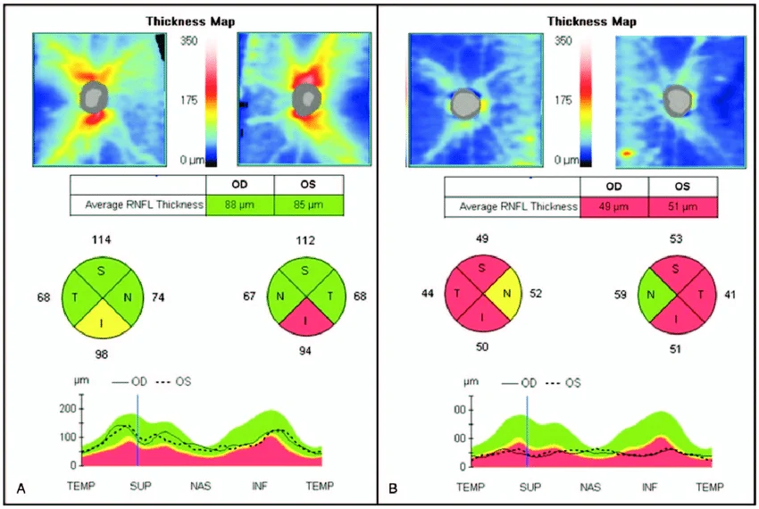
The optic nerve is the cable that carries all visual information from your eye to your brain. When this nerve is damaged — as happens in glaucoma — vision is lost permanently. OCT RNFL scanning allows us to measure the thickness of the nerve fibre layer surrounding the optic nerve with remarkable precision, detecting damage that is completely invisible in any standard eye examination.
OCT stands for Optical Coherence Tomography. It is an advanced imaging technology that uses light waves — not radiation — to produce highly detailed cross-sectional images of the layers of the retina. The RNFL scan specifically focuses on the Retinal Nerve Fiber Layer — the thin layer of nerve fibres that surrounds the optic nerve head. By measuring the thickness of this layer at multiple points around the optic nerve, we can detect thinning that indicates damage, often years before any visual field loss becomes apparent.
Dilating eye drops may be used to widen your pupils for a clearer scan, though many modern OCT machines can perform the scan without dilation.
You rest your chin on the support and look at a target inside the machine. You simply need to keep your eye as still as possible — the scan itself is completely automatic.
A beam of light scans across the retina around the optic nerve head. The machine takes hundreds of measurements in seconds, building a detailed thickness map of the nerve fibre layer.
A colour-coded map appears immediately, showing which areas of the nerve fibre layer are within normal range and which show thinning. Your specialist reviews this map alongside your clinical history and other test results.
Completely. OCT uses light — not radiation — and is entirely non-invasive. The scan takes only a few minutes per eye, is painless, and produces no aftereffects. It can be repeated as often as needed to monitor your condition over time.
Need eye care? Contact us today.
East side of BRB Hospital, 57/E Panthapath, Dhaka-1215
Call Now