
Retina Glaucoma Center & Superspecialty Eye Hospital › Services › VEP (Visual Evoked Potential)
A painless, non-invasive test that measures how visual signals travel from the eyes to the brain. It helps detect optic nerve disorders and other vision pathway problems.
Our specialists are available 6 days a week for in-person and virtual consultations.
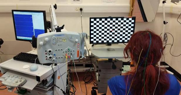
Vision is not just about the eye — it is about the entire pathway from the retina through the optic nerve to the visual cortex at the back of the brain. When something goes wrong anywhere along this pathway, vision is affected in ways that standard eye examinations may not be able to explain. VEP — Visual Evoked Potential — is a specialised test that measures how quickly and how well the visual signal travels from the eye all the way to the brain, allowing us to identify problems anywhere along this entire pathway.
VEP is an electrophysiological test that records the electrical response of the visual cortex — the part of the brain that processes vision — in response to visual stimuli such as a flickering checkerboard pattern. Small electrodes are placed on the scalp over the visual cortex at the back of the head. As you watch the visual stimulus, the brain’s response is recorded. The timing and amplitude of this response reveal how efficiently the visual signal is being transmitted along the entire optic pathway from eye to brain.
Small, flat electrodes are attached to your scalp at the back of the head and at reference points on the forehead and ear. These are held in place with a conductive gel and are completely painless and non-invasive.
You sit comfortably in front of a screen showing a reversing checkerboard pattern. One eye is covered at a time so each eye’s pathway can be tested separately. You simply watch the screen — no active participation is needed beyond keeping your eye open and focused.
The electrodes record the tiny electrical signals generated by your visual cortex each time the checkerboard pattern reverses. Hundreds of responses are averaged together to produce a clear, reliable waveform that shows the timing and strength of your visual cortex’s response.
Your specialist analyses the recorded waveforms, looking at the latency — how long the signal takes to reach the brain — and the amplitude of the response. A delayed or reduced response indicates a problem somewhere along the visual pathway and helps guide the diagnosis.
Need eye care? Contact us today.
East side of BRB Hospital, 57/E Panthapath, Dhaka-1215
Call Now